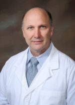
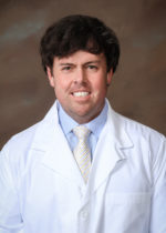
Greenwood Leflore Hospital can boast of one of the finest teams of Orthopedic Surgeons to be found in Mississippi, with Dr. Asa Bennett and Dr. Jay Culpepper standing ready to evaluate and treat everything from broken toes to total hip replacements.
The reputation of Greenwood Orthopedic Clinic has established it as one of Mississippi’s premier bone and joint practices, with its current staff building on the rich legacy created by Dr. Fred Sandifer and Dr. Bill Anderson in the 1970s. Those two surgeons, both graduates of the world-renowned Campbell Clinic training program in Memphis, set the standard for quality care that has been maintained well into the 21st century.
Dr. Asa Bennett joined Greenwood Orthopedic Clinic in 2001. Dr Bennett is a 1995 graduate of the University of Mississippi School of Medicine who went on to complete a General Surgery internship in Chattanooga and an Orthopedic residency at UMMC Jackson. He is also Board Certified and a member of the American Academy of Orthopedic Surgeons and Arthroscopy Association of North America.
Dr. Jay Culpepper a native of Macon, Georgia joined the team at Greenwood Orthopedic Clinic in July 2015. Culpepper completed his medical degree at the University of Mississippi Medical Center and an orthopedic residency at LSU Health in Shreveport, Louisiana.
At Greenwood Orthopedic Clinic, these doctors are well-equipped to handle both chronic degenerative disorders and unexpected injuries. Both have a keen interest in sports medicine and stay busy with the treatment of high school, college and professional athletes as well as that of weekend amateurs. From Friday night sprained ankles to Monday morning golf game aches, Drs. Bennett and Culpepper are dedicated to getting their patients back on the field or court.
Those with a more sedate lifestyle will find Greenwood Orthopedic Clinic accessible and friendly, ready to handle the natural changes of aging in hips, knees and shoulders as well as less common challenges such as rheumatoid arthritis and scoliosis. Whether the patient is headed for surgery or the physical therapist, the orthopedist will be there to guide the treatment plan from beginning to end.
Scope of orthopedic specialty by joint:
Physicians
A hinge-like joint which connects the large upper leg bone (femur) to the larger lower leg bone (tibia) through the kneecap (patella) and a group of cartilage, tendons and ligaments.
Learn more on healthpages.org.
Common pathology: Osteoarthritis, cartilage damage, tears of the meniscus or ligaments
Evaluation: Physical examination, X-rays, MRI or CT scans and examination of extracted joint fluid
Treatment: Surgical and non-surgical options, depending on specific condition and severity
| Non-surgical: |
Surgical: |
| Physical therapy and exercise programs, weight loss, prescription or non-prescription medications or joint injections with steroids or lubricating fluids. |
This may range from minimally invasive procedures to total joint replacement.
Arthroscopy: One or several small incisions are made in the knee to allow insertion of a lighted arthroscope and instruments to repair or remove damaged tissue. Common arthroscopic procedures include meniscus removal or repair, ligament repair or graft, cartilage shaving or removal and patellar smoothing or release.
Open knee surgery: This allows for fuller exposure of the joint in certain cases.
Knee replacement: If damage or deterioriation of the knee is too extensive for repair, a partial or total knee replacement may be necessary. |
A ball-and-socket joint which connects the shoulder blade (scapula) to the large upper arm bone (humerus). The collar bone (clavicle) is another essential element of the shoulder’s function. The rotator cuff is a group of muscles and tendons which connect the shoulder bones and move the joint. The bursa is a fluid-filled sac that cushions the rotator cuff. The labrum is a ring of cartilage within the shoulder joint.
Learn more on healthpages.org.
Common pathology: Osteoarthritis, rotator cuff tears, stretched capsule, labrum tear, fractures and dislocations.
Evaluation: Physical examination, X-rays, MRI or CT scans and examination of extracted joint fluid.
Treatment: Surgical and non-surgical options, depending on specific conditions and severity
| Non-surgical: |
Surgical: |
| Physical therapy and exercise, prescription or non-prescription medications, joint injections.
|
Arthroscopic: Most shoulder problems will involve minimally-invasive arthroscopic procedures, where a small incision allows the insertion of a lighted instrument can be used to visualize the problem. Removal of inflamed tissue, rotator cuff repair, labrum repair and reattachment and removal of bone spurs or loose cartilage may be accomplished without the necessity of open shoulder surgery.
Learn more on nlm.nih.gov.
Open shoulder surgery: More complicated shoulder injuries or degenerative changes may require exposing the entire joint for surgical repair.
Shoulder replacement: In severe cases, the ball of the humerus and the socket of the scapula are replaced with metal and plastic components.
Learn more on WebMD. |
The hands are a complicated network of bones, muscles, tendons and ligaments which work in harmony to allow complex movements essential to daily activities of life.
Common pathology: Osteoarthritis, rheumatoid arthritis, tenosynovitis, contractures, trigger finger, cysts, fractures and dislocations.
Evaluation: Physical examination, X-rays, MRI or CT scans and joint fluid examination.
Treatment: Surgical and non-surgical options, depending on specific condition and severity
| Non-surgical: |
Surgical: |
| Occupational or physical therapy, strengthening and mobility exercises, splints or braces, prescription or non-prescription medications. |
Depending on the nature of the problem, hand surgery may involve a simple incision to access an affected tendon or bone or may require more extensive open hand surgery to repair multiple bones, tendons or ligaments. |
The hips are ball-and-socket joints which unite the pelvis to the large bone in the upper leg (femur). The head of the femur, shaped like a ball, fits snugly into the pelvic depression called the acetabulum, allowing the leg to move forward, backward and side-to-side.
Learn more on healthpages.org.
Common pathology: Osteoarthritis, rheumatoid arthritis, aseptic necrosis, fractures and dislocation
Evaluation: Physical examination, X-rays, MRI or CT scans and joint fluid examination
Treatment: Surgical and non-surgical options, depending on specific condition and severity
| Non-surgical: |
Surgical: |
| Physical therapy, weight loss, anti-inflammatory medications by drugs or by injections. |
Fractures: Hip fractures can result from trauma and/or weakness in the femur from osteoporosis (thinning of the bone). Such fractures usually require surgical intervention to stabilize the joint and restore proper function.
Arthroscopic surgery: Small incisions allow the insertion of a lighted scope and instruments to remove bone spurs or loose cartilage within the hip joint.
Hip replacement: This surgery involves the removal of the hip socket (acetabulum) and the ball (femoral head) and their replacement with an artificial joint made of titanium and plastic. This replacement joint ideally mimics the normal function of the body’s damaged hip.
Learn more on healthpages.org. |